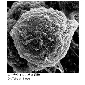
Ebola virus-infected cell (left panel)
Scanning electron micrograph of a cell infected with Zaire ebolavirus.
The cell surface is covered with numerous filamentous Ebola virus particles.
Influenza A virus particles (right panel)
Negative-stain electron micrograph of purified influenza A virus,A/Puerto Rico/8/34 (H1N1).
Haemagglutinin spikes are present on the virion surface. |
The Japanese Society for Virology
|
|